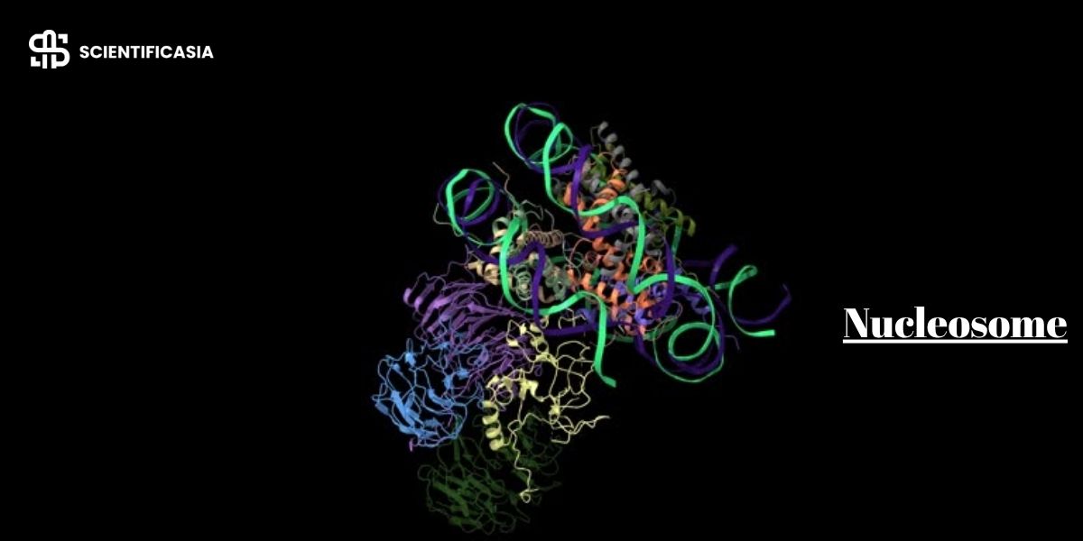
In our body’s tiny cells, the DNA is like a super-long thread, about two meters when stretched out. But it has to fit into a cell nucleus that’s way smaller, less than 10 micrometers across. And not only that, certain parts of the DNA need to be active or inactive at the right times, so the cell can respond to changes around it. It’s like fitting 30 miles of thread into a basketball and being able to pull out the right parts when needed. To do this, from yeast to humans, organisms use something called chromatin, a complex arrangement of their genomes.
The basic piece of this chromatin puzzle is the nucleosome. Imagine it as a building block, repeating every 160 to 240 DNA base pairs along the genome. Each nucleosome is like a spool, holding approximately 145 to 147 base pairs of DNA wrapped around a core of histone proteins. These nucleosome cores are connected by open linker DNA strands to form a continuous chain. Even though the core part is what scientists often refer to as the nucleosome, it’s essential to understand how the entire structure functions to comprehend the intricacies of DNA packing, cellular replication, and architectural survival.
What is a Nucleosome?
The formal nucleosome definition states that a nucleosome serves as DNA’s primary packaging unit in eukaryotes. It is structured like a thread wrapped around a spool made of histone protein octamers, acting as the crucial structural element required to form chromatin and eventually build full chromosomes. To fit within the microscopic boundary of the nucleus, genomic DNA is organized into these units on a massive scale; a single human diploid cell houses around 30 million individual nucleosomes.
Nucleosomes were first glimpsed in 1974 by Don and Ada Olins using an electron microscope, who famously observed that chromatin fibers resembled beads on a string. Their repeating biochemical structure was later elucidated that same year by Roger Kornberg, who isolated the protein interactions. Each nucleosome consists of structured core particles linked by stretches of open DNA, a compact organization that minimizes spatial volume while keeping genetic information completely orderly.
The Core Particle vs. The Chromatosome
When looking at a nucleosome under a high-powered lens, structural biochemists divide it into two primary forms depending on the presence of the external sealing protein:
- The Nucleosome Core Particle: This is the baseline structural unit. It consists strictly of the central eight-part histone spool wrapped by exactly 145 to 147 base pairs of DNA. At this level, the structural spool provides initial compaction but remains open to adjacent blocks.
- The Chromatosome: This represents a more complete packaging state. A chromatosome includes the inner core particle plus an additional stretch of DNA—bringing the total length to about 165 base pairs—which is physically clamped down by a single copy of the external linker protein, Histone H1. The chromatosome acts as a locked, stable structural node that prevents the wrapped genomic thread from uncoiling prematurely.
Detailed Molecular Structure of the Nucleosome
The structure of a nucleosome is a marvel of biochemical engineering, combining double-stranded DNA and a specialized complex of basic histone proteins.
The Histone Core Octamer Assembly
The central spool of the nucleosome is built from an eight-protein complex known as the histone octamer. This core is assembled from two copies each of four highly conserved core histone proteins:
- Two H2A proteins
- Two H2B proteins
- Two H3 proteins
- Two H4 proteins
Structurally, these histones first pair up into heterodimers: H2A binds with H2B, and H3 binds with H4. Two H3-H4 dimers join together to create a central tetramer, which then recruits two separate H2A-H2B dimers to complete the disc-shaped octamer spool.
Electrostatic Binding Mechanics
Because these histone proteins contain exceptionally high concentrations of the basic amino acids lysine and arginine, they carry a strong, permanent positive charge. This positive charge allows them to form highly stable electrostatic bonds with the negatively charged phosphate groups on the outer backbone of the double-stranded DNA molecule.
This interaction is entirely non-sequence-specific, meaning the histone spool can bind and wrap almost any segment of DNA regardless of the genetic code written upon it. However, the physical structure shows a slight preference for flexible, A/T-rich sequences that bend easily around the tight curves of the protein cylinder.
Structural Dimensions and Linker Segments
Each individual disc-shaped nucleosome measures roughly 5.7 nanometers in height and 11 nanometers in diameter. The core DNA wraps around this structure in approximately 1.65 left-handed superhelical loops.
Connecting these bead-like structures is linker DNA, which is usually around 54 base pairs long. This open segment is accompanied by the Histone H1 protein, which sits distinct from the core octamer group, binding where DNA enters and exits the nucleosome to anchor the network. On average, this entire configuration repeats at approximately 200 base pair intervals across the genome.
Primary Biological Functions of the Nucleosome
The nucleosome is not a passive storage spool; it performs critical, active functions necessary to sustain life.
1. Advanced DNA Compaction
The primary function of the nucleosome is to condense massive genomic lengths to fit within the microscopic cell nucleus. To build a full chromosome, this packaging follows a strict structural hierarchy:
- The 10-nanometer Fiber: This is the primary level of compaction where the raw DNA is shortened roughly sevenfold into the basic “beads on a string” format.
- The 30-nanometer Chromatin Fiber: Driven by the chemical interactions of histone tails, the nucleosome chain folds into a thicker fiber using either a helical solenoid or a zigzag pattern.
- Loop Domains and TADs: This thicker fiber forms large loops anchored to a structural scaffold, creating localized functional genomic neighborhoods called Topologically Associating Domains (TADs).
- The Metaphase Chromosome: During cell division, these loops undergo extreme repeated folding to form the dense chromosomes visible under standard microscopes.
2. Physical Genomic Protection
An elegant side benefit of tight structural wrapping is physical preservation. When DNA is wound securely around the histone core, it is sterically shielded from the cellular environment. This means the nucleosome core acts as a protective shield, defending vital genetic sequences from degradation by intracellular nuclease enzymes, while leaving the exposed linker DNA regions vulnerable to regulatory cleavage.
3. Dynamic Control of Gene Expression
Unwinding DNA from this protective histone complex is the first step in reading a gene. Transcription involves copying DNA into ribonucleic acid, or RNA, which then exits the cell nucleus. Some RNA translates into protein in the cytoplasm. Unwinding DNA from its histone complex is the first step in transcription. Specific transcription factors, another group of proteins, bind to particular DNA sequences.
However, these factors can only access the DNA if it is unwound from the nucleosome core. These factors then call upon the DNA polymerase enzyme to start transcription at the right location. DNA polymerase speeds up these reactions, leading to the creation of a complementary RNA strand, also known as mRNA. This process represents just a part of gene expression, a complex dance that controls protein production.
Understanding the Local Chromatin Matrix
To keep genetic studies clear, it helps to understand how an individual nucleosome particle differs from the wider concept of chromatin architecture:
| Genetic Feature | The Nucleosome Particle | Chromatin Architecture |
| Basic Definition | The single repeating structural block made of core DNA wound around a histone spool. | The entire macro-complex of DNA, structural proteins, and RNA forming the chromosome. |
| Primary Elements | Made from exactly eight core histones and roughly 147 base pairs of core DNA. | Contains millions of individual nucleosomes alongside structural scaffolding and linker histones. |
| Functional Focus | Handles local DNA winding and specific chemical modifications on histone tails. | Directs global chromosome folding, loop domains, and large-scale genetic zoning. |
Epigenetic Gene Regulation and the Histone Code
The nucleosome functions as a highly dynamic gatekeeper that directly drives epigenetic gene regulation. When a gene sequence is wrapped tightly inside the spool, the cell machinery cannot read it. The cell manages this through two major biological pathways:
ATP-Dependent Chromatin Remodeling
Cells deploy specialized molecular motor proteins called chromatin remodeling complexes, such as the SWI/SNF family. Using energy from ATP, these complexes physically slide nucleosomes along the DNA strand, alter the structural spacing between spools, or evict them entirely so that transcription factors can easily reach the underlying gene sequences.
Covalent Histone Tail Modifications
Every core histone protein has a flexible, unstructured N-terminal tail that extends outside the core particle. These tails receive specific chemical tags that alter how tightly the nucleosome holds the DNA strand, creating what geneticists call the histone code:
- Histone Acetylation: Enzymes called histone acetyltransferases add acetyl groups to specific lysine residues on the tails. This reduces their positive charge and loosens the bond with the DNA backbone, opening the structure into an accessible, relaxed state called Euchromatin. When histone deacetylases remove these groups, the structure condenses back into an inactive, tightly clustered state called Heterochromatin.
- Histone Methylation: Modifying enzymes add methyl groups to the tails. This does not change the electrostatic charge but serves as a docking node to recruit silencing or activating proteins, depending on the exact amino acid targeted (such as H3K4me3 for activation or H3K27me3 for silencing).
Mitotic Replication and Epigenetic Inheritance
A major challenge for the cell occurs during DNA replication. As the replication machinery moves along the double helix, the tightly bound parental histone spools must be temporarily broken apart and removed so the DNA can be copied.
Immediately behind the advancing replication fork, the cell must assemble new nucleosomes on both daughter strands. To preserve the cell’s identity, old parental histones carrying specific epigenetic tail marks are carefully recycled and distributed alongside newly synthesized histones. This coordinated re-assembly ensures that the delicate histone code is accurately inherited by the next generation of cells, maintaining active and silent genetic zones across cell divisions.
Recent Clinical and Scientific Developments
Cell-Free DNA and Liquid Biopsies
When cells in the body die, their genetic material breaks down and is shed into the bloodstream as cell-free DNA. Because the DNA wrapped tightly inside a nucleosome core is physically shielded from cellular degradation enzymes, the fragments found circulating in blood plasma are highly uniform, typically measuring around 147 base pairs. Modern oncology labs use nucleosome footprinting to map where these particles were positioned, allowing doctors to trace the exact tissue origin of a tumor and monitor cancer changes through a simple blood test.
Circulating Nucleosomes as Cancer Biomarkers
In addition to bare DNA pieces, entire intact nucleosomes carrying tumor-specific histone alterations are shed directly into circulation by cancer cells. Measuring these circulating nucleosomes in plasma samples serves as an effective and sensitive blood-based biomarker for early cancer discovery, showing great success in identifying early colorectal, pancreatic, and lung malignancies.
Advanced Mapping of Fragile Nucleosomes
Using micrococcal nuclease sequencing, scientists digest open linker DNA while keeping nucleosome-covered cores fully intact. This tracking has revealed the existence of fragile nucleosomes, which are unstable or partially unwound histone cores sitting at active promoter sites. Mapping these fragile structures allows researchers to watch exactly how chromatin opens and closes when cells respond to environmental stress or immune triggers.
Summary
Nucleosomes are the key structural units that ensure our lengthy DNA strands can fit neatly inside our cells, serving as the foundational architectural block of eukaryotic life. Far from serving as simple, passive packaging spools, their dynamic structures are essential for maintaining the orderly arrangement of chromosomes, defending genomic material from enzyme degradation, and controlling gene expression. By shifting between tightly coiled heterochromatin and accessible euchromatin, and by maintaining structural integrity during replication, nucleosomes serve as the vital foundations that dictate the daily balance of gene expression and cellular survival.
Update History
Originally Published: August 6, 2023
Current Update: June 14, 2026 — Scientifically reviewed by Muhammad Adeel. Re-structured the entire article architecture into dedicated, high-intent Molecular Structure and Biological Functions sections to match elite competitor formatting rules. Expanded the structural framework by detailing the multi-step assembly of the core histone octamer from dimers to tetramers, alongside non-sequence-specific electrostatic interactions, preferential A/T-rich bending physics, and structural dimensions. Enhanced the functional landscape by integrating macro-compaction pathways (10-nm to 30-nm scales, TADs, and loops), structural nuclease shield dynamics, and mitotic histone recycling pathways during genetic replication.
References
BioNinja. (n.d.). Nucleosomes. IB Biology Syllabus Reference. Retrieved June 14, 2026, from https://ib.bioninja.com.au/nucleosomes/
Kornberg, R. D. (1974). Chromatin structure: A repeating unit of histones and DNA. Science, 184(4139), 868–871. https://www.science.org/doi/10.1126/science.184.4139.868
Luger, K., Mäder, A. W., Richmond, R. K., Sargent, D. F., & Richmond, T. J. (1997). Crystal structure of the nucleosome core particle at 2.8 Å resolution. Nature, 389(6648), 251–260. https://www.nature.com/articles/38444
McGinty, R. K., & Tan, S. (2015). Nucleosome structure and function. Chemical Reviews, 115(6), 2255–2273. ACS Publications. https://pubs.acs.org/doi/10.1021/cr500373h
National Human Genome Research Institute. (2026). Nucleosome. In Talking Glossary of Genomic and Genetic Terms. Genome.gov. Retrieved from https://www.genome.gov/genetics-glossary/Nucleosome
Nucleosome. (2026). In Encyclopedia Britannica. Retrieved from https://www.britannica.com/science/nucleosome
Olins, A. L., & Olins, D. E. (1974). Spheroid chromatin units (v bodies). Science, 183(4122), 330–332. https://www.science.org/doi/10.1126/science.183.4122.330
Snyder, M. W., Kircher, M., et al. (2016). Cell-free DNA architecture identifies cells of origin and variables to transcription factor binding. Cell, 164(1-2), 57–68. ScienceDirect/Elsevier. https://linkinghub.elsevier.com/retrieve/pii/S009286741501569X
Tan, S. (2026). The nucleosome core particle. Tan Laboratory of Structural Biology & Epigenetics at Penn State University. Retrieved from https://sites.psu.edu/tanlab/the-nucleosome/
Wang, Y., Comprehensive Epigenetic Architecture Team, et al. (2026). Single-molecule tracking of parental histone inheritance patterns during replication forks. Nature Communications, 17(1), Article 74448. https://www.nature.com/articles/s41467-026-74448-4
Wikipedia contributors. (2026, May 24). Nucleosome. In Wikipedia, The Free Encyclopedia. Retrieved from https://en.wikipedia.org/wiki/Nucleosome