Cell division is crucial for an organism’s growth and survival. In humans, starting as a single cell in the embryo, divisions lead to over 30 trillion cells in adulthood. This continuous process, known as the cell cycle, includes cytokinesis—the final step.
Cytokinesis begins with cleavage furrow formation during chromosome separation, dividing the cytoplasm between daughter cells. It ends when the bridge connecting them is cut, ensuring successful cell division. Recent research has provided deeper insights into the molecular mechanisms governing this process, including the role of specialized proteins and signaling pathways.
Buckle up, and let’s dive into the article to understand cytokinesis and its impact on growth.
What is Cytokinesis?
Cytokinesis is the last step in the cell cycle, dividing the parent cell’s cytoplasm into two daughter cells through a process known as cytoplasmic division or cell cleavage.
In animals, it starts during anaphase, while in plants, it begins in prophase and concludes in telophase. This results in two daughter cells following mitosis, each with an identical chromosomal set.
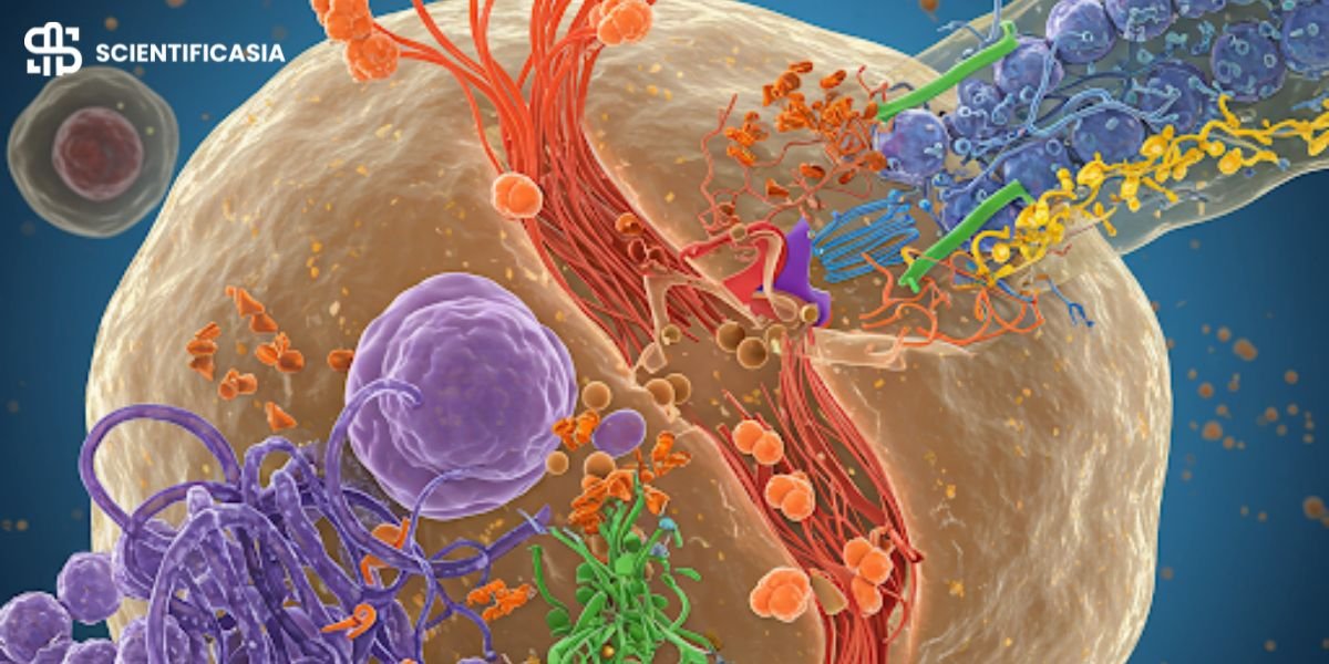
Recent studies show that cytokinesis is regulated by key proteins such as RhoA, which controls the assembly of the contractile ring, and ESCRT-III, which is involved in the final membrane abscission. These proteins ensure precision in cell division, preventing errors that could lead to genetic disorders.
After cytoplasmic division, each new cell is enveloped by a cell membrane, and organelles replicate or synthesize within the cytoplasm. Unlike chromosome replication, cytoplasmic material isn’t doubled, leading to daughter cells being roughly half the size of the parent cell. The daughter cell nuclei remain similar in size due to chromosome replication before mitosis.
Process of Cytokinesis
The cytokinesis process starts alongside mitosis, where a division plane forms at the cell’s center. Microtubules that also create spindle fibers, separating chromosomes in mitosis, shape this plane.
As cytokinesis continues, a contractile ring forms, contracting to make a cleavage furrow. The furrow deepens until the initial cell splits into two. With this, cytokinesis concludes, and the new cells start their cell cycle afresh.
Recent research highlights how the mitotic spindle influences contractile ring positioning through mechanical signals. Errors in this process are linked to diseases like cancer, where cell division becomes uncontrolled.
Cytokinesis also plays a role in creating egg and sperm cells through meiosis. While the procedure mirrors mitosis, a key distinction is that cytokinesis occurs twice in meiosis, forming four new cells, each with just one chromosome copy.
Stages of Cytokinesis
Cytokinesis typically follows the final stages of nuclear division in mitosis and meiosis. It unfolds in four stages: initiation, contraction, membrane insertion, and completion, with variations in animal and plant cells.
Initiation
In the first step, initiation, the goal is to pinpoint where the cleavage furrow forms. The spindle plays a key role, ensuring chromosomes split evenly between nuclei. It also has small structures, astral microtubules, which interact with the membrane and guide actin filament alignment for the upcoming furrow.
Contraction
After identifying the cleavage furrow’s position, actin filaments and proteins like myosin assemble to form the contractile ring. This ring contracts, aiding in the separation of the daughter cells.
Membrane Insertion
Myosin, a motor protein, uses ATP energy to contract the ring further. This deepens the cleavage furrow, gradually forming two distinct cells. Recent findings suggest vesicle trafficking also contributes to membrane expansion during cytokinesis, ensuring proper separation.
Completion
The process concludes as the contractile ring fully divides the cell into two new ones. The final step involves breaking the cell membrane at its narrowest point, allowing the daughter cells to function independently.
Cytokinesis in Animal vs. Plant Cells
Here the difference between cytokinesis in animal and plant cells is described.
| Feature | Animal Cells | Plant Cells |
|---|---|---|
| Division Mechanism | Contractile Ring & Cleavage Furrow | Cell Plate Formation |
| Key Structures | Actin, Myosin, Midbody | Phragmoplast, Vesicles |
| Membrane Contribution | Plasma Membrane Expansion | New Cell Wall Synthesis |
Cytokinesis in Animal Cells
In animals, the cytoplasm tightens until two daughter cells form. A contractile ring under the cell membrane initiates this process. The furrow deepens as actin and myosin contract, eventually leading to cell separation.
The mitotic spindle determines the contractile ring’s position, ensuring chromosome segregation before division. Errors in cytokinesis can lead to abnormal cell division, contributing to tumorigenesis.
Cytokinesis in Plant Cells
In plants, cytokinesis starts with a cell plate forming in the middle, eventually becoming the middle lamella between plant cells. The primary and secondary cell walls develop, separating the daughter cells.
Unlike in animals, the preprophase band determines where the cell plate forms. The phragmoplast, a structure composed of microtubules, helps transport vesicles to build the cell plate. Once fully developed, cellulose is added to strengthen the new cell wall.
Biomedical Applications of Cytokinesis Research
With advancing cell biology research, cytokinesis is being explored in medical and industrial applications:
- Cancer Treatments: Targeting cytokinesis regulators like Aurora kinases for cancer therapy.
- Regenerative Medicine: Cytokinesis studies aid in understanding stem cell division.
- Synthetic Biology: Engineering cell division mechanisms for biomanufacturing.
Proteins Involved in Cytokinesis
Several proteins regulate cytokinesis, including:
- Actinomycin D: Actin forms the contractile ring and is essential for cytokinesis. Disrupting actin filaments halts cell division.
- Myosin: Cytoplasmic myosin II powers the contractile ring’s contraction. Phosphorylation of myosin regulates its movement and function.
- Septins: These proteins assist in linking the plasma membrane to the cytoskeleton, ensuring successful cell division.
- ESCRT-III Complex: This protein complex is crucial in membrane abscission, the final step where daughter cells separate completely.
Conclusion
From the intricate orchestration of contractile rings in animals to the unique cell plate mechanism in plants, cytokinesis ensures the creation of new generations of cells. Advances in molecular biology have revealed how regulatory proteins control this process, offering potential targets for cancer therapies.
The significance of cytokinesis is undeniable. It enables organisms to grow, repair tissues, and reproduce. Understanding this essential process continues to evolve, shedding light on its role in health and disease.


